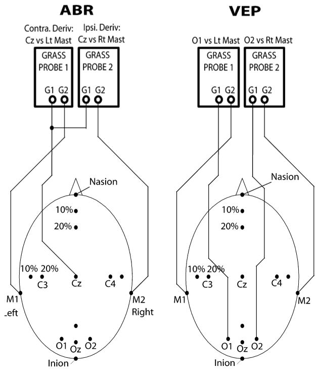
Figure 1.
Auditory brainstem response (ABR) and visual evoked potential (VEP) electrode montage. ABRs were recorded with the active (+) recording electrode at the vertex (Cz). VEPs were recorded with the active (+) electrodes placed at O1 and O2 overlying the left and right occipital cortices, respectively. For both ABRs and VEPs, reference electrodes (−) were placed at the right mastoid (M2) and left mastoid (M1).

