Figure 6.

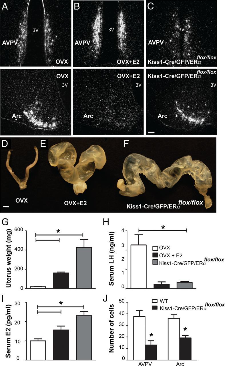
Selective deletion of ERα in Kiss1 neurons disrupts the hypothalamus-pituitary-gonadal axis. A–C, Darkfield photomicrographs of hypothalamic sections showing the distribution of Kiss1 mRNA in the AVPV and Arc nucleus. A, OVX mice showed decreased Kiss1 mRNA in the AVPV and increased Kiss1 mRNA in the Arc. B, OVX estradiol treated (OVX+E2) mice showed increased Kiss1 mRNA in the AVPV and decreased Kiss1 mRNA in the Arc. C, Selective deletion of ERα (Kiss1-Cre/GFP/ERαflox/flox) induced an upregulation of Kiss1 mRNA expression in the Arc. D–F, Representative image showing the uterus of OVX, OVX+E2, and Kiss1-Cre/GFP/ERαflox/flox mice. G, Bar graphs showing the mean uterine weight. Selective deletion of ERα induces a profound enlargement of the uterus. H, Bar graphs demonstrate the LH serum levels from OVX, OVX+E2, and Kiss1-Cre/GFP/ERαflox/flox females. LH levels are elevated in OVX females and are decreased following chronic E2 treatment. Kiss1-Cre/GFP/ERαflox/flox females exhibited similar levels of LH compared with OVX+E2. I, Bar graphs demonstrate the E2 levels from OVX, OVX+E2, and Kiss1-Cre/GFP/ERαflox/flox females. E2 serum levels were significantly higher in OVX+E2 compared with OVX. Kiss1-Cre/GFP/ERαflox/flox females exhibited higher E2 levels compared with OVX or OVX+E2. J, Bar graphs of the average number of Kiss1-Cre/GFP neurons. Kiss1-Cre/GFP/ERαflox/flox females showed a decreased number of AVPV/PeN and Arc Kiss1 neurons compared with wild-type (WT) animals (Kiss1-Cre/GFP). Data are presented as mean ± SEM and *p < 0.05. 3V, Third ventricle. Scale bars: A–C, 50 μm; D–F, 1 mm.
