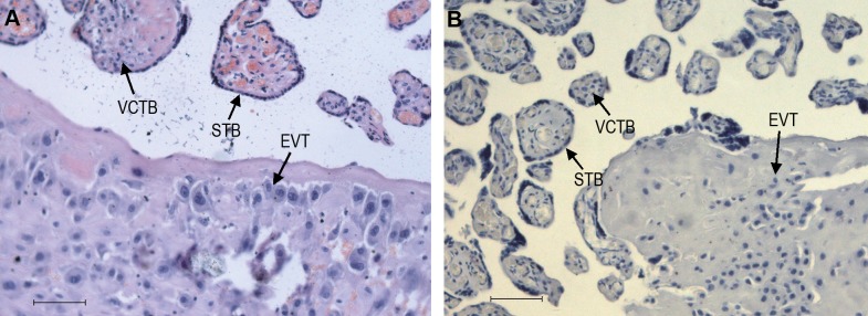
Figure 2.

Hematoxylin and eosin (H&E) staining, and PBS staining of placental tissue sections. A, H&E staining. B, PBS staining as negative control. STB indicates syncytiotrophoblast; VCTB, villous trophoblast; EVT, extravillous trophoblast; PBS, phosphate-buffered saline. Original magnification 200×, scale bar = 40 µm.
