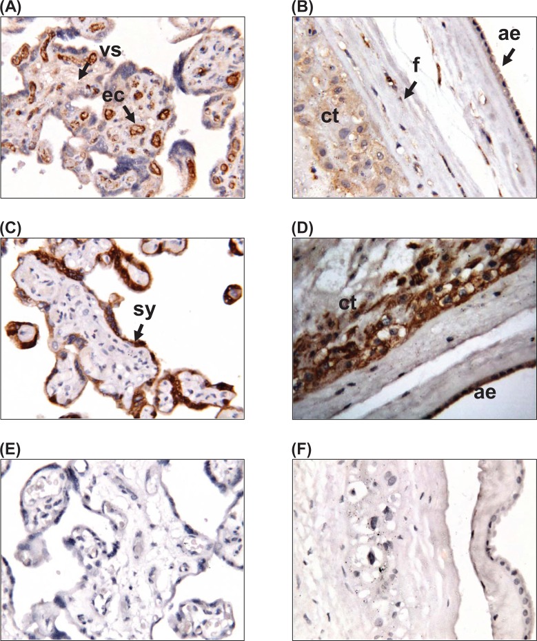
Figure 1.
Localization of apelin and its receptor APJ in human placenta and fetal membranes. Immunohistochemical localization of (A, B) apelin and (C, D) APJ in (A, C) placenta and (B, D) fetal membranes. (E, F) Negative controls for (E) placenta and (F) fetal membranes. Magnification ×250. Sy indicates syncytiotrophoblast cells; cy, cytotrophoblast cells; ec, endothelial cells; ae, amnion epithelium; cl, connective tissue layer; ct, chorionic trophoblast layer; dec, decidua.

