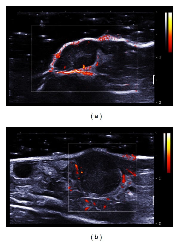
Figure 8.

Visualization of tumour vascularization with Doppler mode (standard Doppler fixed between 0 and 2 m/s). (a) CT26 ectopic tumour and (b) orthotopic CT26 tumour 15 days after tumour implantation. The blood velocity was color coded in scales ranging from 0 to 2 m/s.
