
Figure 2.
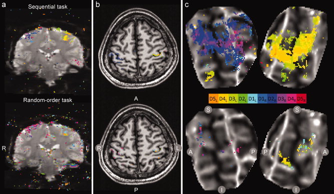
Experiment 1: BOLD activation during sequential and random‐order tasks. Significantly modulated voxels (P < 0.01) in response to cued digit movement are displayed as a color overlay on (a) EPI images, (b) the reference anatomy, and (c) flattened cortical patches. Top: sequential task; bottom: random task. Activation for each voxel was assigned to a digit according to a winner‐take‐all algorithm; significance was estimated for each voxel by permutation analysis. In (a) and (b), data from a single scan are shown (R: right hemisphere; L: left; A: anterior; P: posterior). In (c) data from the average of two scans in a single subject are shown on a flattened cortical patch centered on the central sulcus (dark gray indicates strong negative curvature at the fundus of the sulcus; S: superior; I: inferior). Note the contralateral activation of the hand in the precentral gyrus in both the top and bottom images, but the lack of differentiation between digits in the top image. [Color figure can be viewed in the online issue, which is available at wileyonlinelibrary.com.]