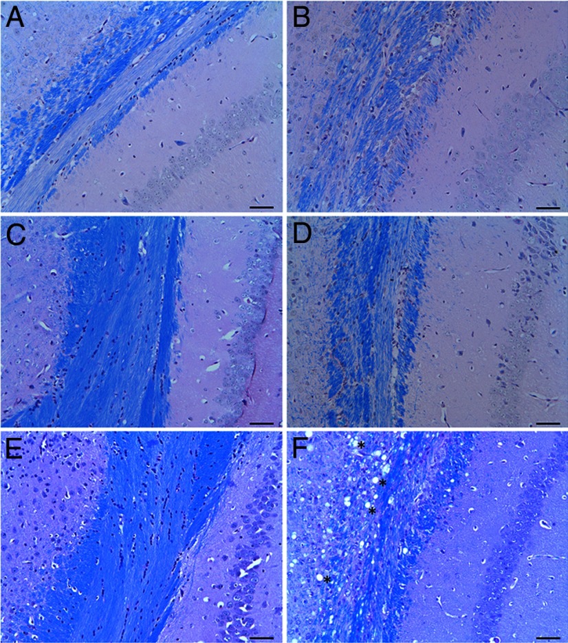
Figure 6.
LFB/PAS-stained coronal sections (5 μm) of the corpus callosum region of Plp1dup compared with wild-type mice. Representative images of brain sections of 1 month (A, B), 3 month (C, D), and 6 month (E, F) wild-type (A, C, E) and Plp1dup (B, D, F) mice. In all pictures, hippocampus is to the right and cerebral cortex to the left. At 3 and 6 months in wild-type mice, LFB staining was homogeneous across the corpus callosum, but in Plp1dup mice at these ages, the staining was uneven and patchy. F, At 6 months, large vacuoles were present in both white and gray matter (*). Plp1dup, n = 3; wild-type, n = 3 at each time point. Scale bar, 50 μm.

