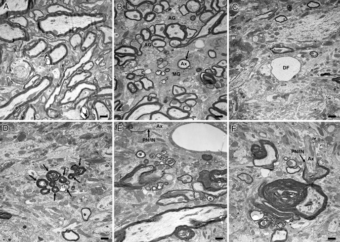
Figure 9.
Electron micrographs of spinal cords from 6 month wild-type and Plp1dup mice. Representative electron micrographs of transition between ventral funiculus and ventral gray matter of wild-type mouse (A) and Plp1dup mouse (B–F) at 6 months of age. Ax, Axon; MG, microglial cell; AG, astrogliosis; DF, large unmyelinated axon (degenerating fiber) packed with neurofilaments; PN/IN with arrow, paranode/internode. B, Arrow points to a degenerating axon being engulfed by a microglial cell. D, Arrows point to a cluster of degenerating fibers. F, The axon (Ax) is myelinated on one side of a node but unmyelinated on the other. Scale bar, 1 μm.

