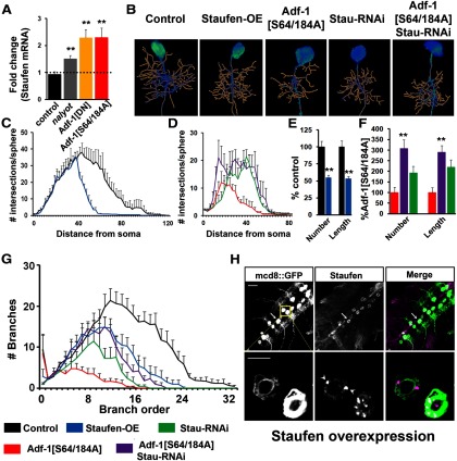
Figure 10.
Adf-1 controls dendrite growth by regulating Staufen transcription. A, qRT-PCR analysis of Staufen expression from adult brains following Adf-1 perturbation. B, Representative RP2 neuron dendrite reconstructions from genotypes that modify Adf-1 and Staufen. C, Volumetric Sholl analysis of Staufen-overexpressing neurons compared with control. D, Volumetric Sholl analysis to test the genetic interaction between Adf-1 and Staufen. E, Quantification of dendrite number and length in Staufen-overexpressing neurons. F, Quantification of dendrite branch number and length for Adf-1–Stau interactions. **p < 0.01. G, Branch-order analysis for Adf-1–Staufen interaction. H, Ventral nerve cords of an EP insertion upstream of Staufen crossed to the RN2-flipout tester GAL4 line stained for mcd8::GFP and Staufen. The bottom row shows close up of RP2 neurons. Arrowheads mark Staufen-positive granules. Scale bars: top row, 50 μm; bottom row, 20 μm.

