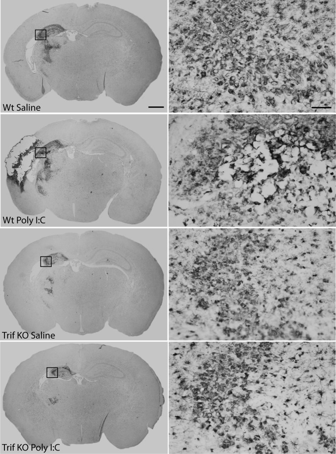
Figure 4.
Inflammatory response after HI. The inflammatory response, as indicated by Iba-1 immunostaining, showed a pattern of amoeboid-like cells in injured brain regions. The degree and distribution of Iba-1 expression appeared to correlate to the degree of brain damage in all experimental groups. The morphology of the Iba-1-positive cells in injured areas did not differ between Poly I:C/HI-treated (B, D) or saline/HI-treated (A, C) animals or between genotypes. Insets represent the area that is depicted at higher magnification. Scale bars: left, 1 mm; right, 5 μm.

