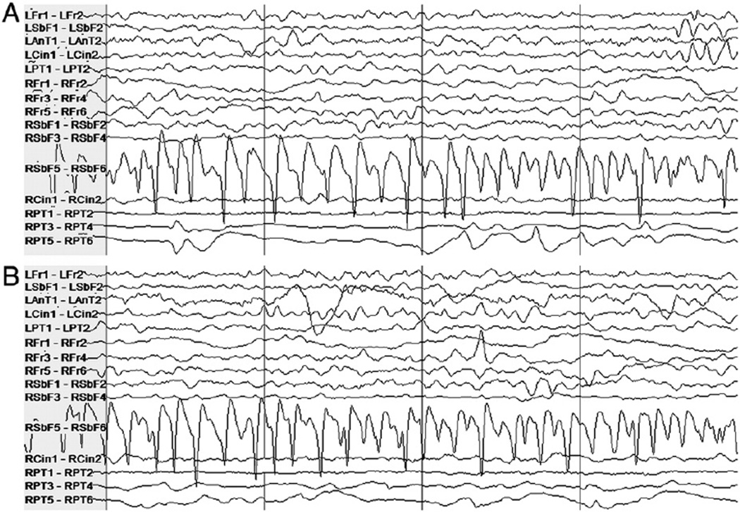
Fig. 4.
Patient 7's intracranial EEG on a subset of electrodes in two epochs showing similar activity, particularly on the right subfrontal channel “RSbF 5–RSbF 6.” (A) Seizure activity in a 4-second epoch beginning approximately 10 seconds after the expert-marked onset. (B) An interictal 4-second epoch during which false detection occurred. This activity was not judged to constitute a seizure by the clinicians.

