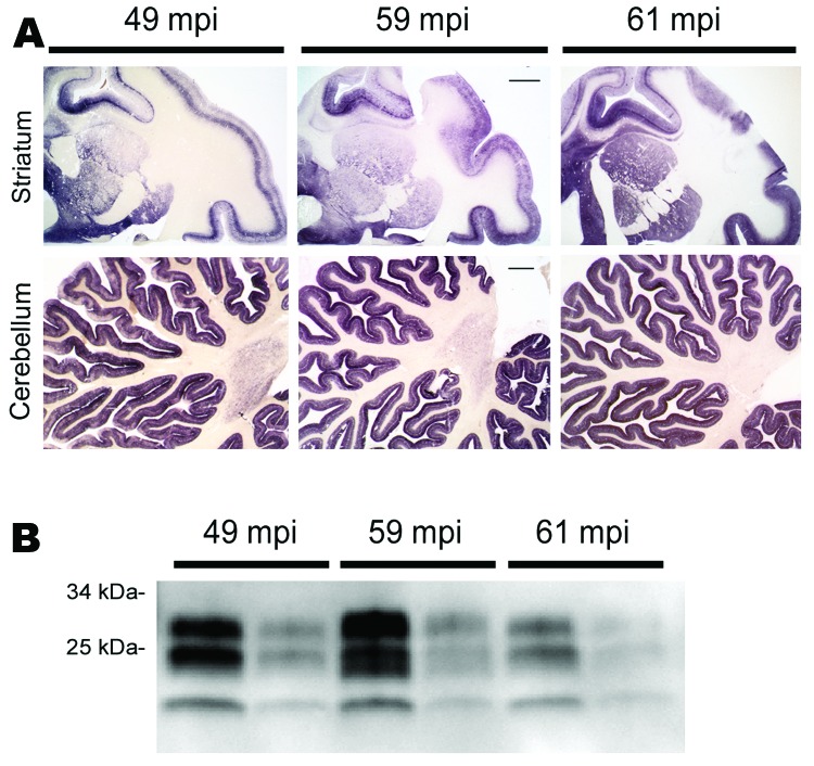
Figure 1.
PrPSc distribution and content in brain of bovine spongiform encephalopathy (BSE)–infected rhesus macaques. A) Paraffin-embedded tissue blot of striatum and cerebellum show a typical BSE-like deposition pattern of PrPSc with no differences between individual BSE-diseased monkeys at 49, 59, and 61 months postinoculation (mpi). Scale bars = 1 mm. B) Western blot analysis for PrPSc in brain of BSE-infected monkeys with incubation times of 49, 59, and 61 mpi. PrPSc-type is as expected for BSE prions, and no major differences in PrPSc load were detected. All samples were proteinase K–digested; loading amount was 0.5 and 0.1 mg fresh wet tissue for each sample

