
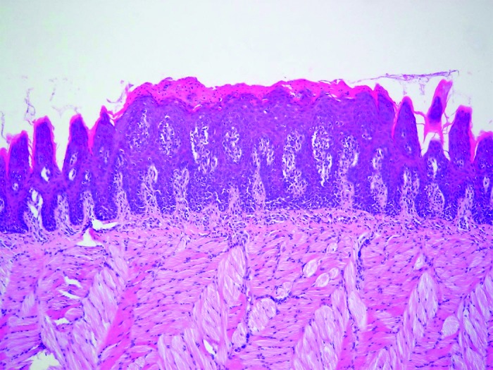
Figure 5. Sagittal section of rat tongue dorsum showing tissue lesion characterized by epithelial hyperplasia, disorganized basal layer, exocytosis, spongiosis, loss of filiform papillae, and hyperparakeratosis. HE: 100×.
Official websites use .gov
A
.gov website belongs to an official
government organization in the United States.
Secure .gov websites use HTTPS
A lock (
) or https:// means you've safely
connected to the .gov website. Share sensitive
information only on official, secure websites.
Figure 5. Sagittal section of rat tongue dorsum showing tissue lesion characterized by epithelial hyperplasia, disorganized basal layer, exocytosis, spongiosis, loss of filiform papillae, and hyperparakeratosis. HE: 100×.