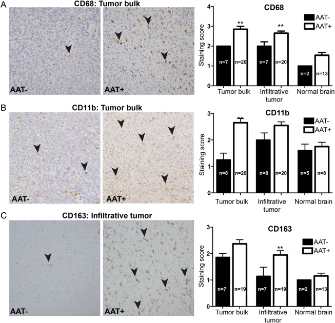
Fig. 1.
TAM measurement by immunohistochemistry. TAMs were morphologically identified and then semiquantitatively scored on the basis of the following markers: CD68, CD11b, and CD163. The staining score was defined as follows: 0 (0% of cells stained), 1 (1%–10% of cells stained), 2 (11%–50% cells stained), 3 (51%–90% cells stained), and 4 (>90% cells stained). Microscopically, an increase in the number of TAMs was seen in AAT+ patients. (A) An increase in CD68+ TAMs (black arrowheads) was seen in tumor bulk (P < .01) and infiltrative tumor (P < .05) in AAT+ patients than in AAT– patients. (B) An increase in CD11b+ TAMs (black arrowheads) was seen in tumor bulk (P < .01) of AAT+ patients than in that of AAT– patients. (C) An increase in CD163+ TAMs (black arrowheads) was seen in infiltrating tumor (P < .05) in AAT+ patients.

