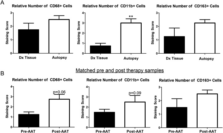
Fig. 3.
TAM analysis in matched samples. (A) When available, the initial diagnostic tissue of AAT+ patients was examined and compared with their autopsy tissue. A relative increase in CD68+, CD11b+, and CD163+ TAMs were seen in autopsy, as compared with their initial diagnostic tissue, with significance reached for CD11b+ cells (P < .01). (B) A separate cohort of patients treated with antiangiogenic therapy was examined. Pretreatment tissue was compared with posttreatment tissue. Although statistical significance was not reached, likely because of the limited number of samples (n = 4), there was a trend for an increase in CD68+ TAMs (P = .06) and CD11b+ cells (P = .09).

