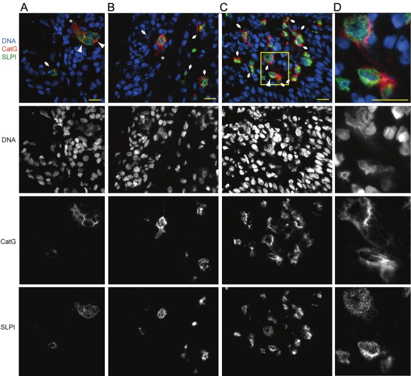
Figure 1.

CatG- and SLPIcontaining NETs are present in psoriatic skin. Fluorescence microscopy images of lesional skin stained for CatG (red), SLPI (green) and DNA (blue). Netting cells (A-C), enlarged in (D), were identified by diffuse nuclei (demonstrating a lower-intensity Hoechst staining pattern), intracellular chromatin dispersion or extracellular fibrous DNA deposits. Cells with dispersed chromatin that co-stained with CatG and SLPI are shown by arrows. Neutrophils undergoing CatG release that is not associated with DNA and/or SLPI are shown by asterisks. Arrowheads highlight CatGSLPI-DNA co-localization at the cell surface. Data are from one donor and are representative of at least three donors. Scale bar = 10 µm.
