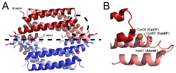
Fig. 1.

Crystal structure of Mycobacterium tuberculosis CsoR15 (A) and Cu(I) binding site (B). CsoRs feature a tetrameric bundle as a “dimer of dimers” displayed as blue and red and separated by a dashed line. There are four Cu(I) binding sites (bronze spheres), two per dimer. In CstR, Cys31 and Cys60′ form cross-links with the corresponding protomer of the dimer.7 Labelled residues are that of M. tuberculosis CsoR and those in parentheses are the analogous residues in S. aureus CstR.
