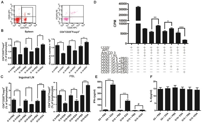
Figure 2. Effects of T. gondii ESA on the proportion and function of CD4+CD25+Foxp3+ T cells at different stages of pregnancy.
All animals were killed at G18, and their spleens, inguinal LN, and PBL were obtained. Lymphocytes from these tissues were prepared and pooled as described in Materials and Methods. The cells were stained with CD4-FITC, CD25-APC and PE-Foxp3 Abs, respectively, and analyzed by flow cytometry. (A)Representative dot plots illustrating the regions and gating for the capture of cell phenotype data and intracellular Foxp3 expression. (B) Percentages and absolute number of CD4+CD25+Foxp3+-cells from spleens. (C) Percentages of CD4+CD25+Foxp3+-cells from inguinal LN, and PBL. (D) Responder CD4+CD25– T cells (1×10⁵/well) from naive mice were cultured with naive, irradiated APC (1×10⁵ cells/well) and CD4+CD25+T cells (5×10⁴ cells/well) harvested from mice with PBS or T. gondii ESA injection at G5, G10, G15, respectively. (E and F) The serum levels of IFN-γ and IL-4 in pregnant mice injected with T. gondii ESA by ELISA. Data represent means ± SD from groups of seven mice assayed individually. Statistical differences between groups are shown as follows: * p<0.05; ** p<0.01; *** p<0.001; # p>0.05.

