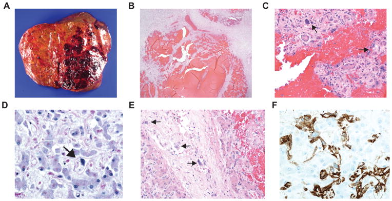
Figure 1. Hepatic Angiosarcoma in Patient with Dyskeratosis Congenita.

A, macroscopic photograph showing a large hemorrhagic lesion in the left lobe with several smaller lesions in the right lobe of the liver. B, photomicrograph of liver, demonstrating peliotic changes with resultant blood sequestration caused by the tumor (hematoxylin-eosin, original magnification x2). C, photomicrograph of liver, demonstrating enlarged, hyperchromatic tumor cells (black arrows) admixed with normal liver cells (hematoxylin-eosin, original magnification x20). D, photomicrograph of right lobe of the liver (away from macroscopic lesions), demonstrating diffuse infiltration of tumor cells (black arrow) throughout the liver (hematoxylin-eosin, original magnification x40). E, photomicrograph of liver, demonstrating tumor cell (black arrows) infiltration into the wall of an interlobular blood vessel (hematoxylin-eosin, original magnification x20). F, photomicrograph of liver, the tumor cells are immunopositive for CD31, an endothelial marker, consistent with hepatic angiosarcoma (CD31 immunohistochemistry, original magnification x40).
