
Figure 7.
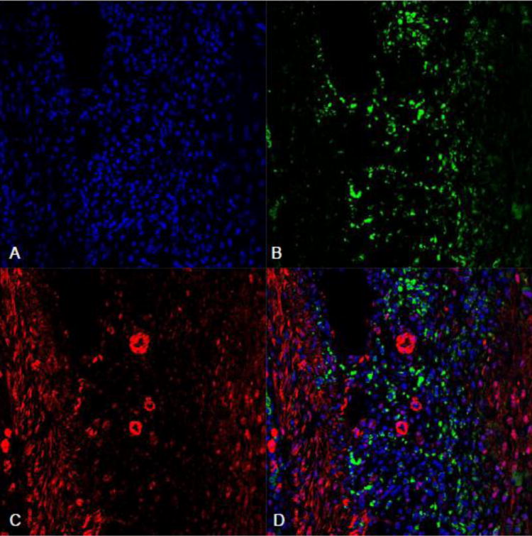
Fluorescent ED-1 and VEGFR-3 double staining in the infarcted myocardium: ED-1+ macrophages (green) were accumulated in the infarcted myocardium at day 7 postMI (panel B). Panel C showed VEGFR-3 staining (red) in the same region. Overlapped images of ED-1 and VEGFR-3 (panel D) showed that VEGFR-3 was negatively expressed in macrophages. Panel A: nuclear dapi staining.