Fig. 3.
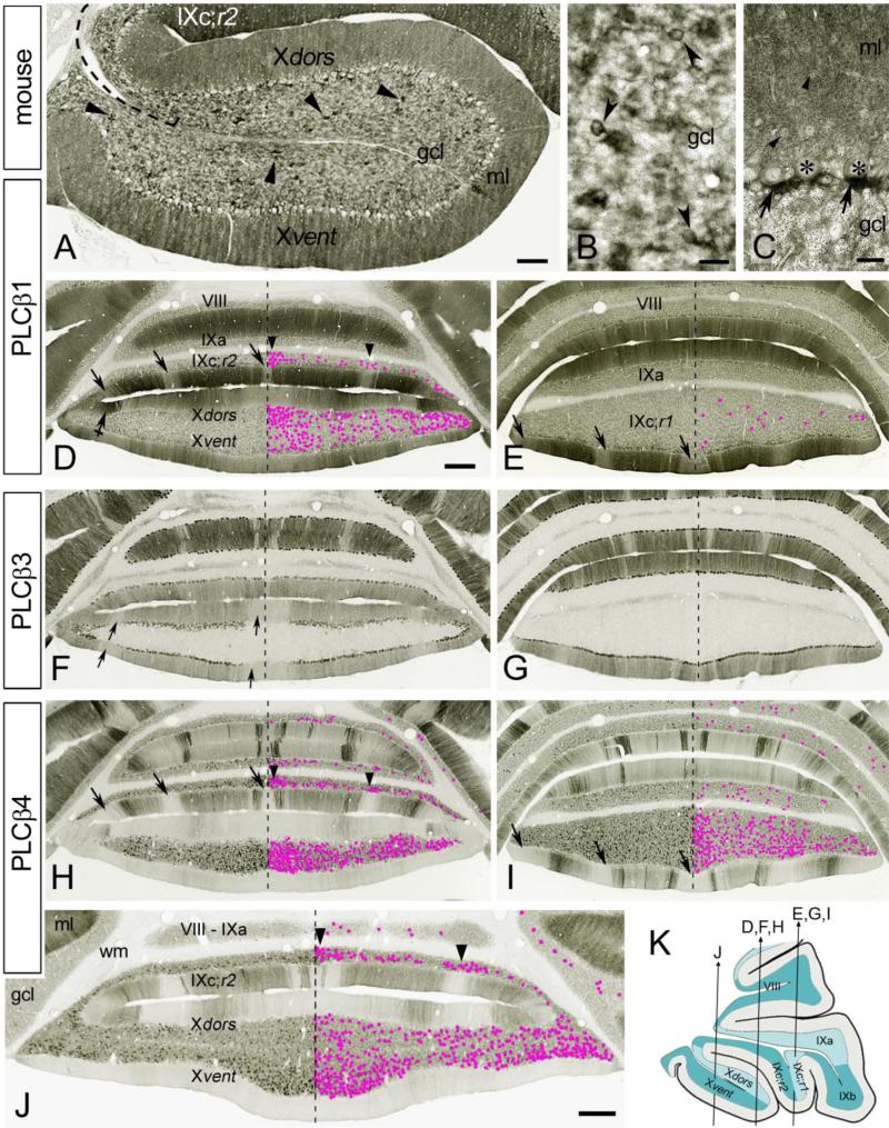
Cryosections of the mouse cerebellum immunostained with antibodies to PLCβ1 R-233 (sc9005) (a-e), PLCβ3 (Frontier Institute) (f, g), and PLCβ4 (h-j). a Parasagittal paraffin section of nodulus shows moderate PLCβ1 staining throughout the gcl. The somata and brushes of UBCs (arrowheads) are noticeably PLCβ1+. Note the moderately immunopositive Purkinje cell bodies with unstained nuclei and the homogeneous immunolabeling of the ml. Dashed line delineate the tz between IX and X. b Representative PLCβ1+ UBCs (arrowheads) from the nodulus; cryosection. c Detail from lobule V shows moderately stained cell bodies of basket/stellate cells (arrowheads) and intensely PLCβ1+ pinceaux (arrows) below the Purkinje cell somata (asterisks); paraffin section. d, e Purkinje arbors in the ml display moderate to intense PLCβ1 immunostaining. Few lighter stained bands (arrows) are noticeable, especially in IXc;r1&r2. The nodulus and IXc;r2 contain the highest densities of PLCβ1+ UBCs (d), especially the midline portion of IXc;r2. Few PLCβ1+ UBCs are present in IXc;r1 (e), while PLCβ1+ UBCs are absent in other lobules included in sections (d, e). Bands with high densities of PLCβ1+ UBCs (arrowheads) are present in IXc;r2. Crossed arrow point to intensely labeled lateral stripe in Xdors. f, g PLCβ3-immunostaining shows on/off Purkinje cell band in the ml. The PLCβ3 and PLCβ4 show reciprocal immunostaining in the Purkinje cells bands, with exception in nodulus and IXc;r2. UBCs are PLCβ3-. In nodulus three PLCβ3- Purkinje cell stripes are present, in both Xdors and Xvent (arrows); one wide stripe at midline and 2 lateral stripes (one on each side). h-j The ml shows on/off bands of PLCβ4–immunoreactivity in Purkinje arbors. In the nodulus many Purkinje cells are PLCβ4-, especially in the Xvent (h, j). PLCβ4+ UBCs are present at high concentration in nodulus and IXc;r1&r2 (h-j), and especially in median portions of the Xvent and IXc;r2 (h, j). PLCβ4+ UBCs are also present in other lobules (h-j). The distribution of PLCβ4+ UBCs does not seem in register with Purkinje cell on/off bands, with the exception of IXc;r2 (h, j), in which high density UBC bands are situated approximately beneath PLCβ4- Purkinje cell bands (arrowheads). In IXc;r1&r2 the PLCβ4- bands (arrows) are situated at the same position as are the moderately stained PLCβ1+ bands in the adjacent sections (arrows in d, e). k Schematic drawing showing the approximate planes of the coronal cerebellar sections in d-j. Panels e, f, and h show adjacent sections, as do panels e, g, and i. Dashed line in panels d-j indicates the cortical midline. PLCβ1+ UBCs (d, e) and PLCβ4+ UBCs (h-j) in the gcl are marked by magenta dots. Scale bars a 200 μm,b, c 20 μm, d, j 0.5 mm (d applies to d-i)

