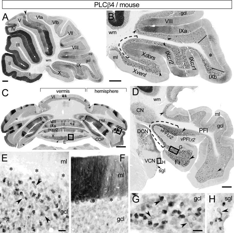
Fig. 5.
PLCβ4-immunoreactivity in mouse cerebellum visualized by DAB chromogen. a Sagittal midvermal section illustrates the distributions of PLCβ4-immunostained Purkinje cells and UBCs. At this particular vermal level the Purkinje cells in anterior lobules I-V and around the primary fissure (arrowhead) show the most immunolabeling, while they are either moderately labeled or unstained in the posterior lobules VIb and VII-X. The UBC-rich nodulus, uvula and lingula show distinct PLCβ4 immunoreactivity in the gcl. b Enlargement of the caudal cerebellum illustrates the distribution of PLCβ4+ UBC (arrowheads) in the gcl. Nodulus, IXc;r1&2 (delineated by white dashed line), and IXb contain the highest densities of PLCβ4+ UBCs. Tz IX to X is indicated by black dashed line. Few PLCβ4+ UBCs are present in IXa and VIII. Asterisk marks a distinct field of uvula (between IXa and IXb, delineated with solid lines) that is devoid of UBCs. Most of the Purkinje cells in nodulus are PLCβ4-. c Coronal section of the posterior cerebellum displays the parasagittal stripes of the PLCβ4+ Purkinje cells and the distribution of PLCβ4+ UBC (arrowheads). The nodulus and uvula show the highest density of PLCβ4+ UBCs, additionally, scattered PLCβ4+ UBCs are distributed in other vermal and hemispheral lobules. d Fl, PFl, lateral CN, and adjacent brain stem in coronal section. High densities of PLCβ4+ UBCs (arrowheads) occur in Fl and vPFl;r1&2, especially at the transition zone between the two structures (black dashed line). In CN small neurons are distinctly immunolabeled, while the neuropil shows moderate staining. Within the cochlear nuclear complex, PLCβ4+ UBCs occur in the DCN and the sgl (arrow), but not in the VCN. e Enlarged boxed area from panel c shows a dense PLCβ4+ UBC population (arrowheads) in nodulus. Asterisks mark PLCβ4- Purkinje cell bodies. f Enlarged boxed area from panel c shows adjacent, intensely and moderately immunolabeled Purkinje cell dendrites. Asterisks indicate Purkinje cell bodies. g, h Enlarged boxed areas from panel d show PLCβ4+ UBCs (arrowheads) in Fl (g) and in sgl (h). The cerebellar wm is unstained (a, b, d). Scale bars a-d 200 μm, e, f 25 μm, g, h 20 μm

