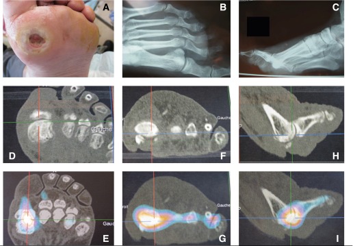
Figure 2.

Chronic ulcer of the first metatarsal head. A: Photograph. B and C: Plain radiographs. D, F, and H: Three-dimensional X-ray tomographic images. E, G, and I: Corresponding 67Ga SPECT/CT images. The gallium accumulation marks the best puncture place.
