
Figure 7.
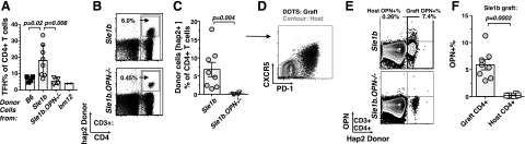
OPN supports the sustained survival of donor CD4+ and TFH cells in bm12 recipients. CD4+ T cells were analyzed 4 wk post-transfer into bm12 recipients, as described in Fig. 5. A) Flow cytometric comparison of TFH cells isolated from bm12 recipients after the transfer of B6, Sle1b, or Sle1b.OPN−/−. Gray bars indicate mean values of data points; error bars indicate se. B) Tracking of donor Sle1b and Sle1b.OPN−/− CD4+ T cells by flow cytometry employing a Slamf-haplotype 2 specific antibody. Bottom panel represents staining of the spleen of the recipient with the highest percentage of engraftment. C) Statistical analysis of the quantitation of Slamf-haplotype 2 donor cells by the Mann-Whitney nonparametric, 2-tailed test. D) Donor Sle1b CD4+ T cells are CXCR5+ PD-1+, as judged by flow cytometry. E) Intracellular flow cytometry staining of OPN in Sle1b donor (Slamf-haplotype 2) and recipient CD4+ T cells. Bottom dot plot represents a spleen with the highest percentage of engraftment. F) Statistical analysis of E.