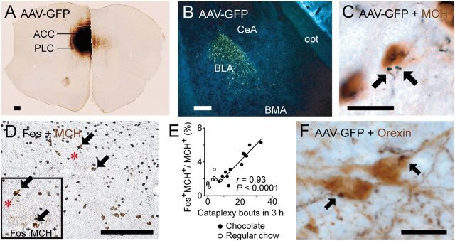
Figure 6.
The ACC/PLC innervates brain regions that may regulate cataplexy. A, B, Many ACC/PLC neurons express GFP immunoreactivity after injection of the anterograde tracer AAV-GFP and they heavily innervate the BLA. C, GFP-labeled axons (black) also closely appose MCH neurons (brown). D, E, Double labeling for Fos (black) and MCH (brown) shows that Fos expression in the MCH neurons is positively correlated with the number of cataplexy bouts (chocolate: n = 12; regular chow: n = 8). F, GFP-labeled axons from the ACC/PLC (black) closely appose orexin neurons (brown). Scale bars, 200 μm (A, B,D) and 20 μm (C,F). BMA indicates anterior basomedial nucleus of the amygdala; opt, optic tract.

