Figure 1.

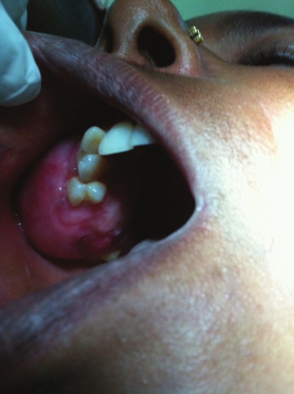
Intraoral photograph showing smooth lobulated swelling, 2 × 5 cm in diameter extending from maxillary first premolar to first molar on right side and posteriorly to the maxillary tuberosity area

Intraoral photograph showing smooth lobulated swelling, 2 × 5 cm in diameter extending from maxillary first premolar to first molar on right side and posteriorly to the maxillary tuberosity area