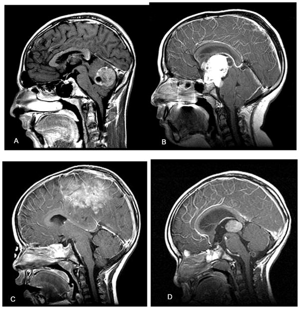
Figure 1.
Sagittal MRIs of pediatric brain tumors in different brain regions. (A) Posterior fossa tumor in 14-year-old male with brief history of nausea, vomiting, and headache. At surgery, the lesion proved to be a medulloblastoma. (B) Midline suprasellar mass lesion with invagination into 3rd ventricle in 10-year-old female with progressive visual loss and headaches. This tumor was a craniopharyngioma. (C) Parasagittal tumor in 2-year-old female with brief history of seizures and leg weakness. This tumor was an ependymoblastoma. (D) Posterior third ventricular tumor in 2-year-old male with a history of vomiting and headaches. The lesion was a pineoblastoma.

