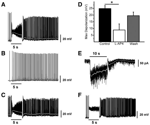
Figure 6. Synaptically-evoked light response of M3 cells in WT mice.
(A–C) Example of typical synaptic light response of an M3 cell recorded in current clamp mode to a 5 s, full-field, bright white light stimulus first in control conditions (A), in the presence of 100 μM L-AP4 (B), and following washout (C). (D) Mean ± SE maximum depolarization evoked by 5 s full-field bright white light stimulus in control (black bar), 100 μM L-AP4 (white bar), and after washout (gray bar). (E) Voltage clamp recording of M3 cell response to 10 s full field, bright, white light stimulus. (F) Example of typical synaptic light response of an M2 cell recorded in current clamp mode to a 5 s, full-field, bright white light stimulus in the absence of synaptic blockers. Gray lines (A–C) represent 0.1 s smoothing of membrane voltage. * p < 0.05 ANOVA.

