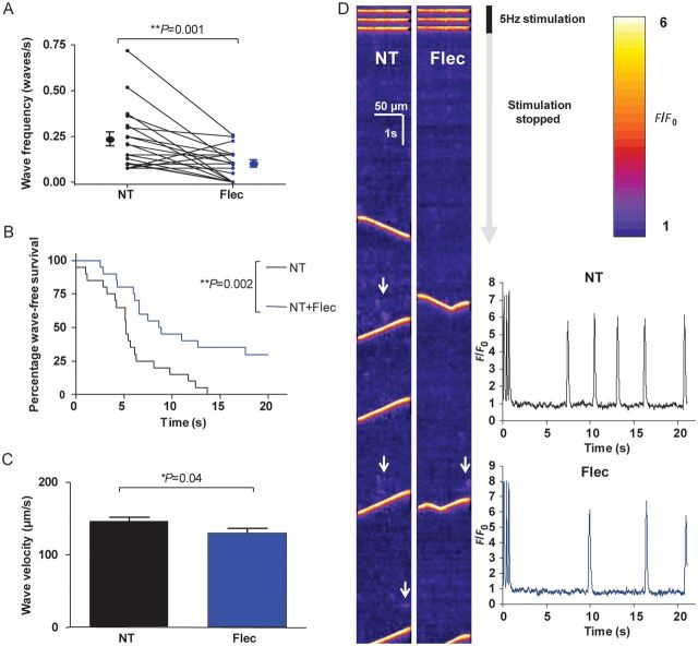
Figure 2.
Effects of 5 µM flecainide on Ca2+ waves. (A) Flecainide was washed on or off via cross-over protocol for 5 min. In the presence of flecainide, wave frequency was significantly reduced (P = 0.001, n = 20 cells). (B) Latency period from the last transient to the first wave is shown in the Kaplan–Meier survival format (i.e. wave-free survival). Cells in the presence of flecainide have an increased wave-free survival period (P = 0.002 by log-rank test, n = 20 cells). (C) Wave velocity is reduced in the presence of flecainide (P = 0.04 by Student's t-test, NT: n = 81 waves; flec: n = 36 waves from 20 cells). (D) Representative line-scans from a cell assessed for waves pre- and post-flecainide application. The end of the 30 s period of 5 Hz stimulation evoking Ca2+ transients can be seen at the top of the scans with subsequent quiescent phase during which waves are observed. Areas of increased spark activity prior to waves are highlighted with white arrows and are more prominent in the absence of flecainide. Inset: line-scans converted into F/F0 plots—reduction of wave frequency and increased latency is apparent.

