
Figure 1.
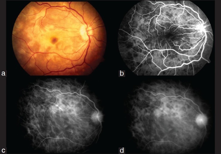
Active Serpiginous choroiditis (a) Color fundus photograph of the right eye showing presence of yellowish white subretinal confluent lesions radiating away from the disc sparing the fovea, (b) Transit phase FFA showing presence of confluent hypofluorescent lesions radiating away from the disc not involving the fovea with staining around these lesions, (c) Early phase ICGA showing presence of numerous confluent hypofluorescent lesions around the disc and surrounding the fovea, (d) Persistence of the hypofluorescent lesions with involvement of fovea with areas of loss of choriocapillaris on ICGA