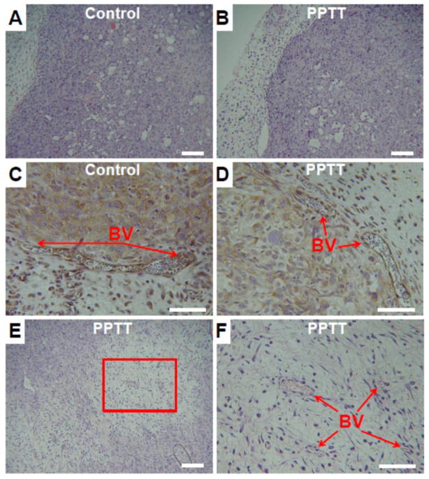
Fig. 6. Histology of control and PPTT treated tumors.
No differences between the tumor periphery of both control and PPTT treated tumors were observed (A–B, 10x objective, scale bar = 100 μm). IHC staining of blood vessels (BV) in the periphery did not provide evidence of damage in either group (C–D, 20x objective, scale bar = 50 μm). The center of tumors treated with PPTT had evidence of cell and tissue damage most likely due to excessive heating in this region (E, 10x objective, scale bar = 100 μm). A higher resolution view of this area shows the presence of capillary blood vessels which appear viable despite surrounding damage (F, 20x objective, scale bar = 50 μm).

