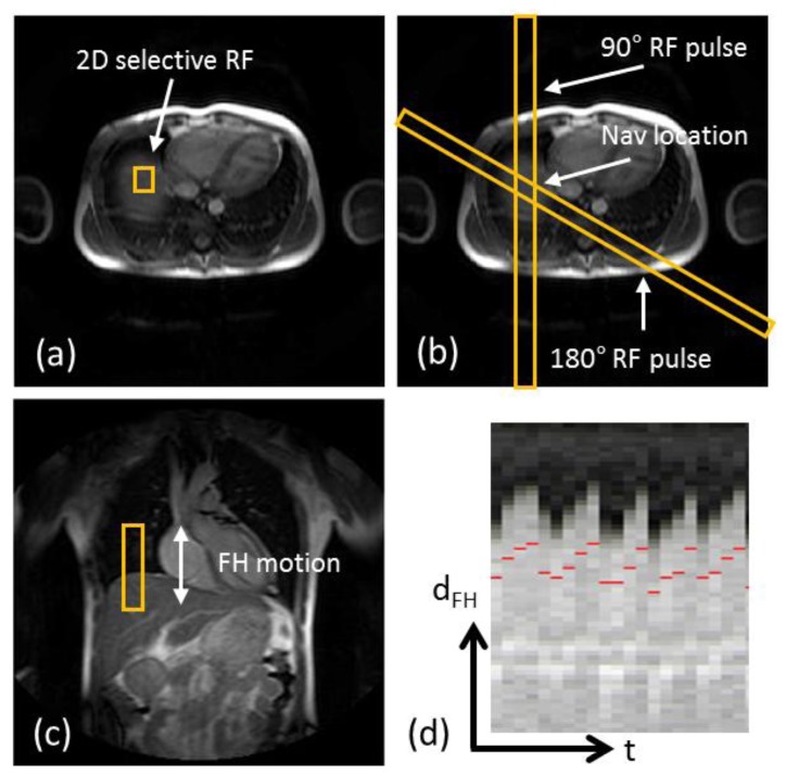
Figure 3.
Scan planning of d1D NAV in axial plane using either a 2D selective RF “pencil beam” excitation (a) or a spin-echo approach with obliquely aligned 90° excitation and 180° refocusing pulses (b). The d1D NAV positioned in the coronal plane, on the dome of the right hemi-diaphragm with the readout in foot-head (FH) direction (c). The d1D NAV signal (d) clearly captures the displacement of the lung-liver interface along the FH direction (dFH) over time (t).

