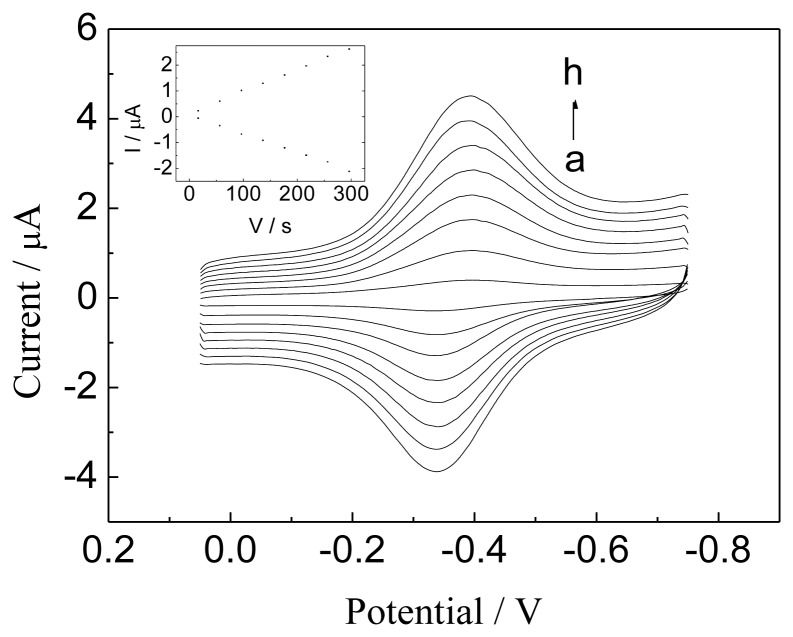
Figure 5.
CVs of the Hb/GNPs-GR-SDS/BPG electrode in N2-saturated PBS (pH 7.0) at different scan rates. Scan rate (from a to h): 0.06, 0.1, 0.14, 0.18, 0.22, 0.24, and 0.3 V·s−1. The inset plot shows the linear dependence of anodic and cathodic peak currents vs. scan rate in the range 0.06-0.3 V·s−1.

