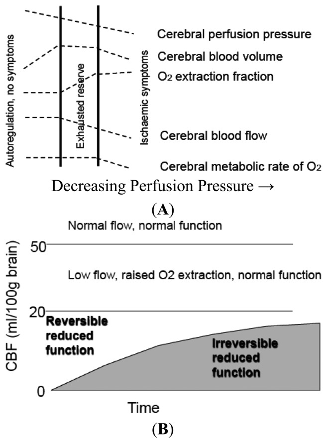
Figure 1.
(A) Cerebral Blood Flow and Metabolic Thresholds. With falling Cerebral Perfusion Pressure (CPP) as occurs distal to a cerebral artery occlusion, intracranial arteries dilate to maintain CBF- a process termed autoregulation. This results in an increase in Cerebral Blood Volume. When vasodilation is maximal, further falls in CPP result in a fall in CBF and results in increase in Oxygen Extraction Fraction (OEF) to maintain tissue oxygenation. When OEF is maximal further falls in CPP lead to reduction in Cerebral Metabolic Rate for Oxygen utilization (CMRO2). (B) The combined effects of residual CBF and duration of ischemia on reversibility of neuronal dysfunction during focal ischemia. The gray shaded region outlines the limits of severity and duration of ischemia, distinguishing tissue “not at risk” from functionally impaired tissue. Schematics are drawn from concepts attributed to Baron and Heiss et al [25,26].

