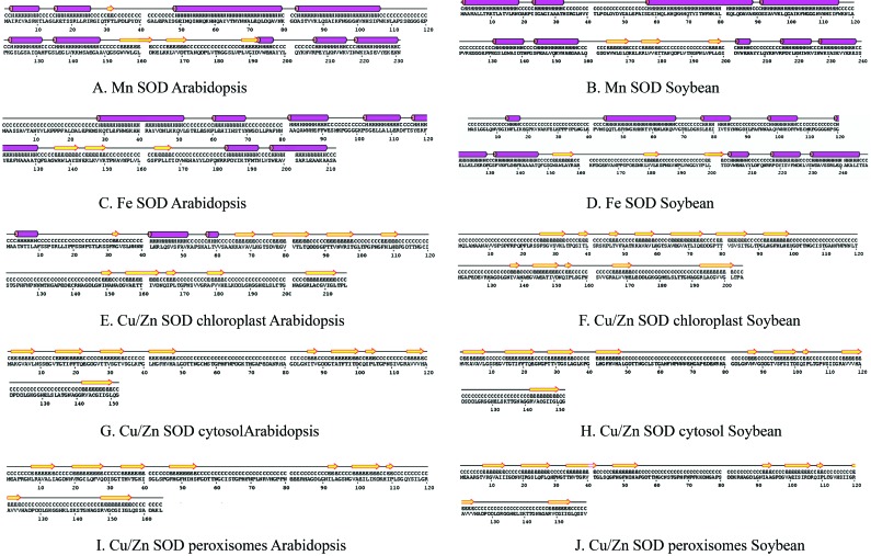
Figure 3.
Alignment of the secondary structures of soybean SOD with Arabidopsis SOD homologues. Identical residues are placed adjacent to facilitate comparison. The predicted secondary structure is shown for each aligned peptide: yellow arrows indicate β-sheets and pink cylinders indicate α-helices.

