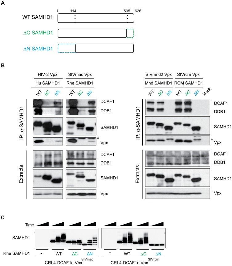
Figure 4. N- and C-terminal binding Vpx proteins degrade SAMHD1 through a conserved mechanism.
(A) Schematic representation of wild type (WT) SAMHD1, C-terminally truncated SAMHD1 (ΔC, shown in green), and N-terminally truncated SAMHD1 (ΔN, shown in blue). Amino acid numbers of the truncations are shown, with dotted line indicating the truncated region of SAMHD1. (B) HA-SAMHD1 (WT, ΔC, and ΔN) were transiently co-expressed in 293T cells with the autologous FLAG-Vpx and immunoprecipitated from whole cell extracts with anti-HA resin. HA-SAMHD1, FLAG-Vpx, DCAF, and DDB1 were detected in immune complexes (top panels) or extracts (bottom panels) by western blotting. * denotes the antibody light-chain. (C) In vitro ubiquitylation of WT, ΔC, and ΔN rhesus SAMHD1, in the presence or absence of SIVmac Vpx (left panel) or SIVrcm Vpx (right panel). SAMHD1 and Vpx were incubated with Cul4, DCAF1c, RBX1, UBA1 (an E1 enzyme), UbcH5b (an E2 enzyme) and FLAG-tagged ubiquitin for increasing time (0, 15, 30 min), and ubiquitylation of each SAMHD1 construct was analyzed by western blotting.

