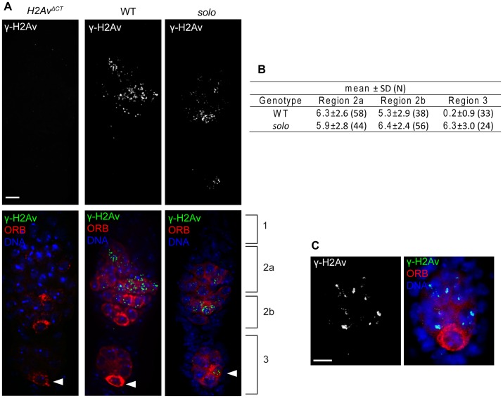
Figure 6. solo mutations cause a transient delay in DSB repair.
Pro-oocytes and oocytes from solo mutant (soloZ2-3534/Df(2L)A267) and WT females were identified and staged by ORB staining and relative positions within germarium. γ-H2Av was stained by anti- γ-H2Av antibody and DNA was visualized with DAPI. γ-H2Av foci from pro-nurse cells were not scored. Scale bars: 5 µm. (A) γ-H2Av staining in germaria. Left panel shows absence of antibody staining in P{w+H2AvΔCTXC}; l(3)H2Av810 females in which the only expressed histone H2Av subunits are deficient for serine-137 and are not phosphorylated in response to DSBs [66]. Center and right panels show antibody staining in WT and solo germaria. γ-H2Av staining is absent in region 3 oocyte in WT but present in region 3 oocyte in solo (arrowheads). (B) Average number of γ-H2Av foci per nucleus in pro-oocytes and oocytes at different stages. N is the number of nuclei scored. (C) Absence of γ-H2Av staining in the oocyte nucleus of a stage 2 solo egg chamber. Egg chamber was from the same solo ovariole from which the germarium shown in panel A was taken.

