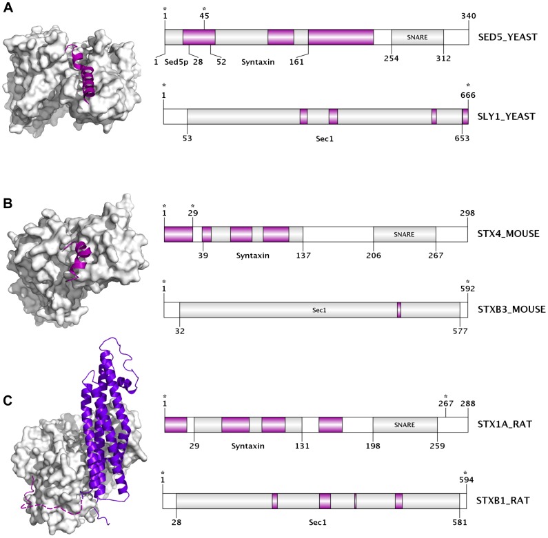
Figure 2. Interactions between the disordered N-terminal tails of SNARE proteins and their globular SM protein partners.
Three PDB structures are presented showing the interaction between distinct pairs of the syntaxin-family SNARE proteins and their regulatory SM-proteins, an interaction that has been shown to positively regulate the SNARE complex assembly. The N-terminal of the SNARE partner is predicted to be mostly disordered (more than 50% of its residues) in the unbound form in all cases. (A) Interaction between the yeast syntaxin-family SNARE Sed5 N-terminal region, and SM protein Sly1 (PDB: 1MQS). (B) Interaction between the N-terminal tail of syntaxin-4 and syntaxin-binding protein 3 from mouse (PDB: 2PJX). (C) Interaction between syntaxin-1A (structure lacking the C-terminal transmembrane region) and syntaxin-binding protein 1 from rat (PDB: 3C98). Each interaction pair is represented by a PDB structure (left) and a domain map of the entire protein chain for both partners (right). The upper domain map corresponds to the SNARE protein, while the bottom one to the SM partner. In the structures, the disordered SNARE N-terminal tails are represented with cartoon style (magenta) while the partner molecule is in surface representation (white). In panel C, the remaining part of syntaxin-1A, which is not part of the disordered N-terminal tail, is coloured purple-blue, and those disordered residues of the N-terminal missing from the X-ray structure (10–26) are represented by a dashed-line. Names and lengths are provided for each protein in the corresponding domain map. Names and locations of their known Pfam domains (predicted by the PfamScan method) are also indicated. Regions predicted to be disordered (length of at least 3 consecutive residues) by IUPred are coloured in magenta, while the ordered segments are white (if not predicted to be part of a Pfam domain) or light-gray (if they are). Regions present in the PDB structures are marked by stars.

