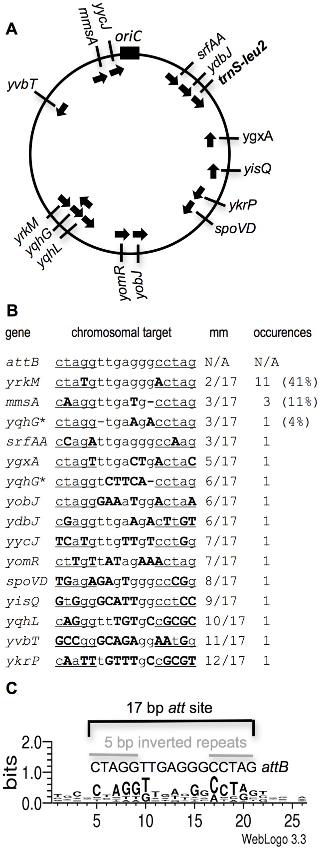
Figure 2. Map and DNA sequence of the primary and 15 secondary integration sites for ICEBs1.

A. Approximate position of the primary and 15 secondary ICEBs1 integration sites on the B. subtilis chromosome. The circle represents the B. subtilis chromosome with the origin of replication (oriC) indicated by the black rectangle at the top. The slash marks represent the approximate location of the ICEBs1 insertion site. The name of the gene near which (ygxA) or into which (all other locations) ICEBs1 inserted is indicated on the outside of the circle. The arrows on the inside of the circle indicate the direction of ICEBs1 replication for each insertion. trnS-leu2 (in bold) contains the primary ICEBs1 integration site attB. B. DNA sequence of the primary and 15 secondary integration sites. The gene name is indicated on the left, followed by the DNA sequence (chromosomal target). The primary attachment site (attB) is a 17 bp sequence with 5 bp inverted repeats (underlined) separated by a 7 bp spacer. Mismatches from attB are indicated in bold, capital letters. “mm” indicates the number of mismatches from the primary 17 bp attB. “occurrences” indicates the number of independent times an insertion in each site was identified. Percentages of the total (27) are indicated in parenthesis. The * next to yqhG indicates that two different ICEBs1 insertions were isolated in this gene, once in each orientation. C. Sequence logo of the ICEBs1 secondary attachment sites. Using Weblogo 3.3 [21], we generated a consensus motif of the 26 bases surrounding the insertion site of the 15 secondary insertion sites for ICEBs1. For comparison, the primary attachment site for ICEBs1 is a 17 bp region with 5 bp inverted repeats and a 7 bp spacer region in the middle [10]. The size of each nucleotide corresponds to the frequency with which that nucleotide was observed in that position in the secondary attachment sites.
