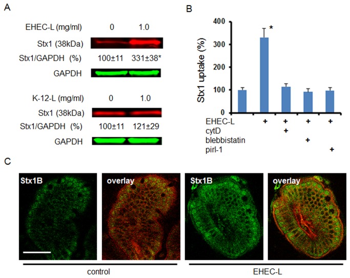
Figure 5.
EHEC-L stimulate Stx1 MPC in mouse ileum.
(A) Representative IB and quantitative representations of data show that EHEC-L significantly increases Stx1 uptake in mouse enterocytes compared to tissue treated with K-12-L (n ≥ 6 animals per each experimental condition; * - significant compared to the control (p = 0.03)). (B) EHEC-L-induced Stx1 uptake in mouse intestine was reduced to the control level in the presence of inhibitors of MPC including cytD (n = 3 mice), blebbistatin (n = 4 mice) or pirl-1 (n = 4 mice). (C) Representative multiphoton optical section either through control sample of ileal tissue exposed to Stx1 only or tissue treated with EHEC-L plus Stx1 shows substantial increase in Stx1 fluorescence inside the enterocytes. In both panels: plasma membranes – red by tdTomato, Stx1-488 – green; bar -50 µm.

