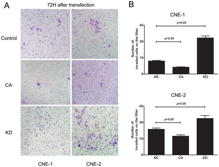
Figure 6. Inhibition of GSK-3β activity enhanced invasion of NPC cells.
(A) Representative photos showing the NPC cell density on the filter after transfection with GSK3β plasmid. Inhibition of GSK-3β by GSK3β-KD transfection enhanced invasion of CNE-1 and CNE-2 cells, whereas activation of GSK-3β by GSK3β-CA transfection suppressed invasion of CNE-1 and CNE-2 cells; (B) Quantitative analyses of the number of invaded cells showed that the invaded cells in GSK3β-KD group increased significantly, whereas those in GSK3β-CA group decreased significantly when compared to the control. Invasion of NPC cells was evaluated by transwell assay after transfection with GSK3β-KD or CA plasmid (2 μg/mL) for 72 h. The data indicate the means (SEM) of 3 independent experiments. NC, normal control; CA: constitutively active GSK-3β plasmid; KD: kinase-dead GSK-3β plasmid.

