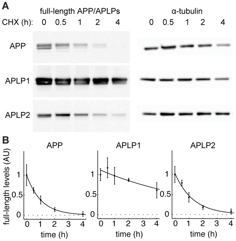
Figure 3. APP and APLP2 have a higher protein turnover than APLP1.
(A) Western blot analysis of HEK cells transfected with C-terminally HA-tagged APP/APLPs after indicated times of protein synthesis inhibition with cycloheximide (CHX). Western blots were probed with anti-HA antibody. Note the strong accumulation of APLP1 as compared to APP and APLP2. (B) APP/APLP full-length levels from A were normalized to α-tubulin. Mean ± SEM of n = 3 are shown for each time point. Data was fitted to exponential functions by the least square approach. R2 (APP) = 0.99; R2 (APLP1) = 0.82; R2 (APLP2) = 0.98.

