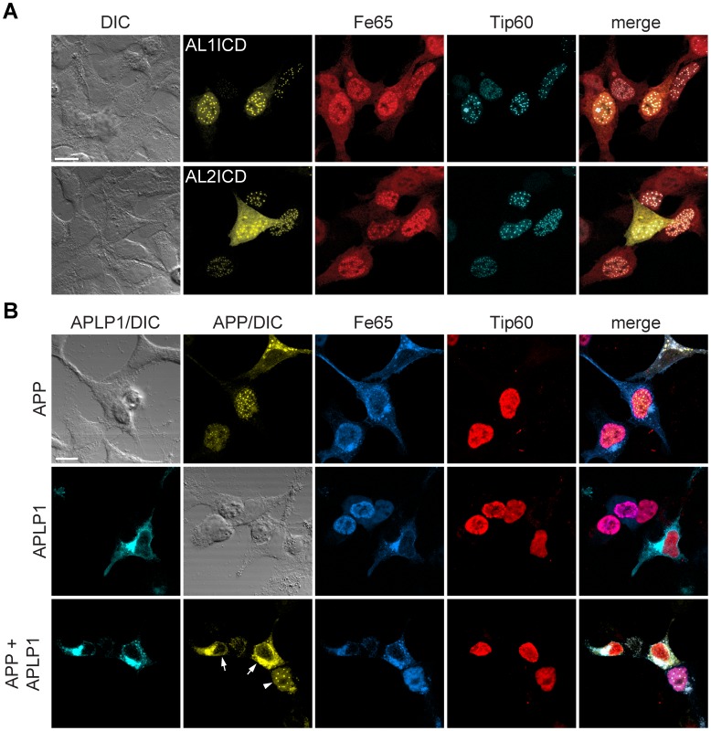
Figure 8. APLP1 expression prevents localization of AICD to AFT complexes.
(A) Confocal fluorescence images of HEK cells transfected with HA-Fe65, CFP-Tip60 and Cit-AL1ICD (top row) or Cit-AL2ICD (bottom row). Note the nuclear localization of Al1ICD to nuclear complexes. (B) Confocal fluorescence images of HEK cells transfected with HA-Fe65, Myc-Tip60 and cotransfected with APP-Cit (top row) APLP1-Cer (middle row) or both (bottom row). Note that AFT complex formation (arrowhead) was ablated in cells expressing APP as well as APLP1 (arrows). Scale bars represent 13 µm.

