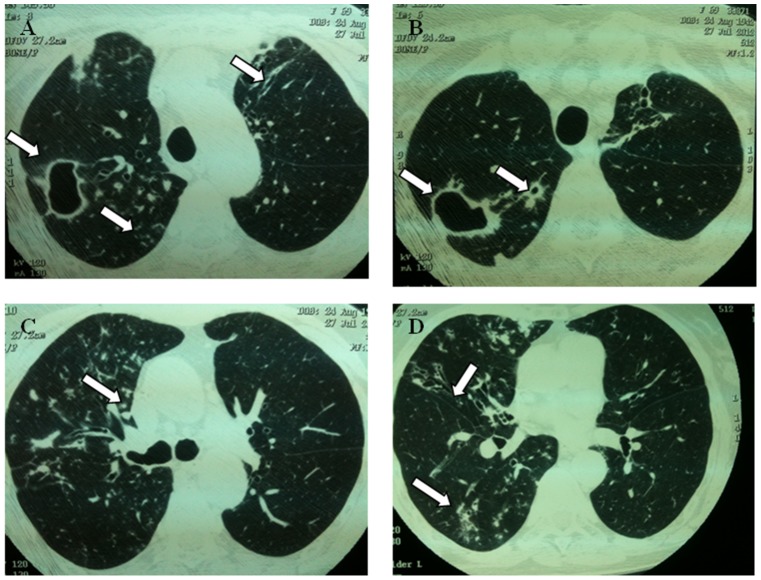
Figure 2. Representative image showing lung damage in a patient with nontuberculous mycobacterial diseases.
A 69-years-old woman with Mycobacterium abscessus pulmonary disease. (A and B) High resolution computed tomography (HRCT) of the chest obtained at level of upper lobes showing multiples cavities in the right upper lobe and centrilobular nodules. It is also possible to observe bronchiectasis in left upper lobe (arrow). (C) Tree-in-bud pattern. (D) Presence of bronchiectasis in middle lobe. Also note centrilobular nodules at right lower lobe.

