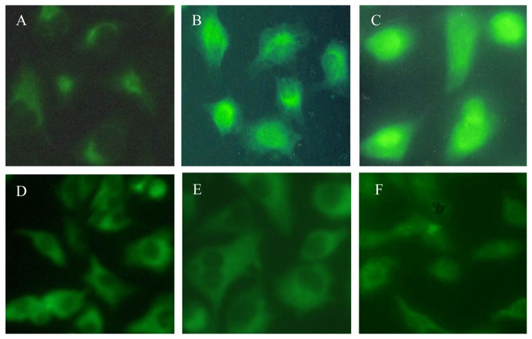
Figure 1. Egr-1 expression and localization in lung epithelial cell line A549.
(A) 0 minute (control cells, grown in medium alone), Egr-1 expression was detected but very weak and located in cytoplasm. (B) 30 minutes, the expression of Egr-1 was increased and partly located in cytoplasm. (C) 60 minutes, robust expression of Egr-1 was located in the nuclear. (D) 120 minutes, medium expression of Egr-1 was located in cytoplasm and nuclear. (E) 240 minutes, less expression of Egr-1 was located in cytoplasm. (F) 480 minutes, Egr-1 expression almost restored to the level of control cells. Images were at ×100 magnification.

