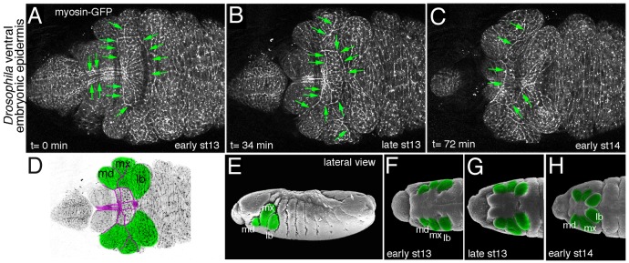
Figure 2. A multitude of actomyosin cables during morphogenesis of the ventral epidermis of the fly embryo. (A-C) Still images of a time-lapse move showing myosin-GFP in the ventral anterior epidermis during major epithelial morphogenetic movement occurring concomitant with the process of head involution (early stage 13 to early stage 14 of embryogenesis). Green arrows point to many actomyosin cables observed during this process. (D) Schematic of late stage 13 epidermis with the maximillar, mandibular and labial segments indicated. Magenta lines trace the actomyosin cables observed at this time point. (E-H) Illustrated scanning electron micrographs of the ventral epidermis corresponding to the images in (A-C) [modified from Flybase (www.flybase.org); Images from F.R. Turner. Personal Communication to Flybase, 1995]. Mandibular, maximillar and labial segments are marked in green. Anterior is to the left in all images, all panels show ventral views apart from (E) which shows a lateral view.
