Fig.1.

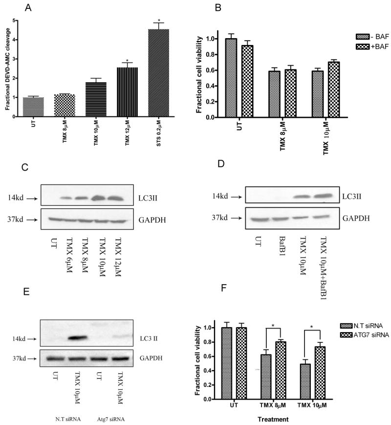
OHT triggers autophagic death in MPNST cells. (A) OHT-treated cells demonstrated a concentration-dependent increase in levels of caspase-3 like activity (48 hours). * indicates p<0.05 relative to untreated cells. However, broad caspase inhibition with BAF (50μM, 1hour pre-treatment) (B) did not provide protection from OHT-induced death (48 hours). Exposure to OHT (48 hours) triggered (C) a concentration-dependent increase in steady state levels of LC3-II, and (D) an increase in autophagic flux. Cells transfected with siRNA against Atg7 demonstrated (E) unchanged levels of LC3-II following OHT treatment (48 hours) and (F) partial protection from OHT-induced cytotoxicity (72 hours). * indicates p<0.05 relative to cells transfected with N.T siRNA.
