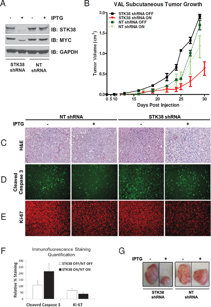
Figure 5. Suppression of STK38 in human B-cell lymphoma can delay tumor progression.
(A) Immunoblot analysis of STK38 protein silencing at 48 hours after IPTG (400 uM) induction of shRNA expression in stable VAL cell lines expressing an IPTG-inducible shRNA against either STK38 or a non-targeted (NT) sequence. (B) Tumor growth of the VAL stable cell lines injected into SCID mice ±SEM. The VAL STK38 shRNA cells, with or without IPTG, are shown as red and black lines, respectively. The VAL NT shRNA cells, with or without IPTG are shown as light and dark green lines, respectively. (C–E) VAL STK38 shRNA cells and NT shRNA cells before and after IPTG induction. (C) H&E staining (D) Immunofluorescence staining for cleaved caspase 3 (E) Immunofluorescence staining for Ki-67. (F) Quantification of the cleaved caspase 3 and Ki-67 staining. Bars represent the percentage of positively stained VAL STK38 shRNA cells with or without IPTG induction normalized to the NT shRNA cells with or without IPTG ±SEM, respectively. (G) Representative tumors excised at Day 25 post-injection.

