
Figure 7.
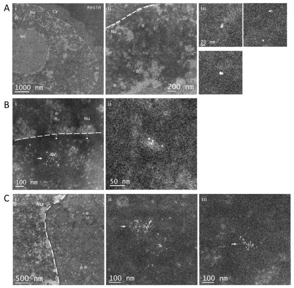
Intracellular imaging of Au(GSH) nanoparticles. (A) (i) STEM imaging allows a clear delineation of the cytoplasm and nucleus in plastic-embedded HeLa cells prepared with no heavy-metal contrast agents. (ii) Higher magnification image of the region marked with an asterisk in (i). (iii) Expanded views of the regions marked with asterisks in (ii). In (iii), small Au(GSH) nanoparticle aggregates are visible inside the nucleus. (B) (i) Au(GSH) nanoparticles both in and outside the nucleus. Nanoparticles outside the nucleus appear mostly clustered together and are likely to be inside endosomes (arrow). Nanoparticles marked with arrowheads appear isolated and are within 5–10 nm of the nuclear membrane, suggesting they are free in the cytosol. (ii) Expanded view of the region in the nucleus marked with an asterisk in (i). (C) Au(GSH) nanoparticles outside the nucleus, probably within endosomes (arrow). (ii) and (iii) are higher magnification images of the regions marked with one and two asterisks in (i), respectively. Nu, nucleus; No, nucleolus; Cy, cytoplasm. The dashed lines mark the boundary between the nucleus and cytoplasm.