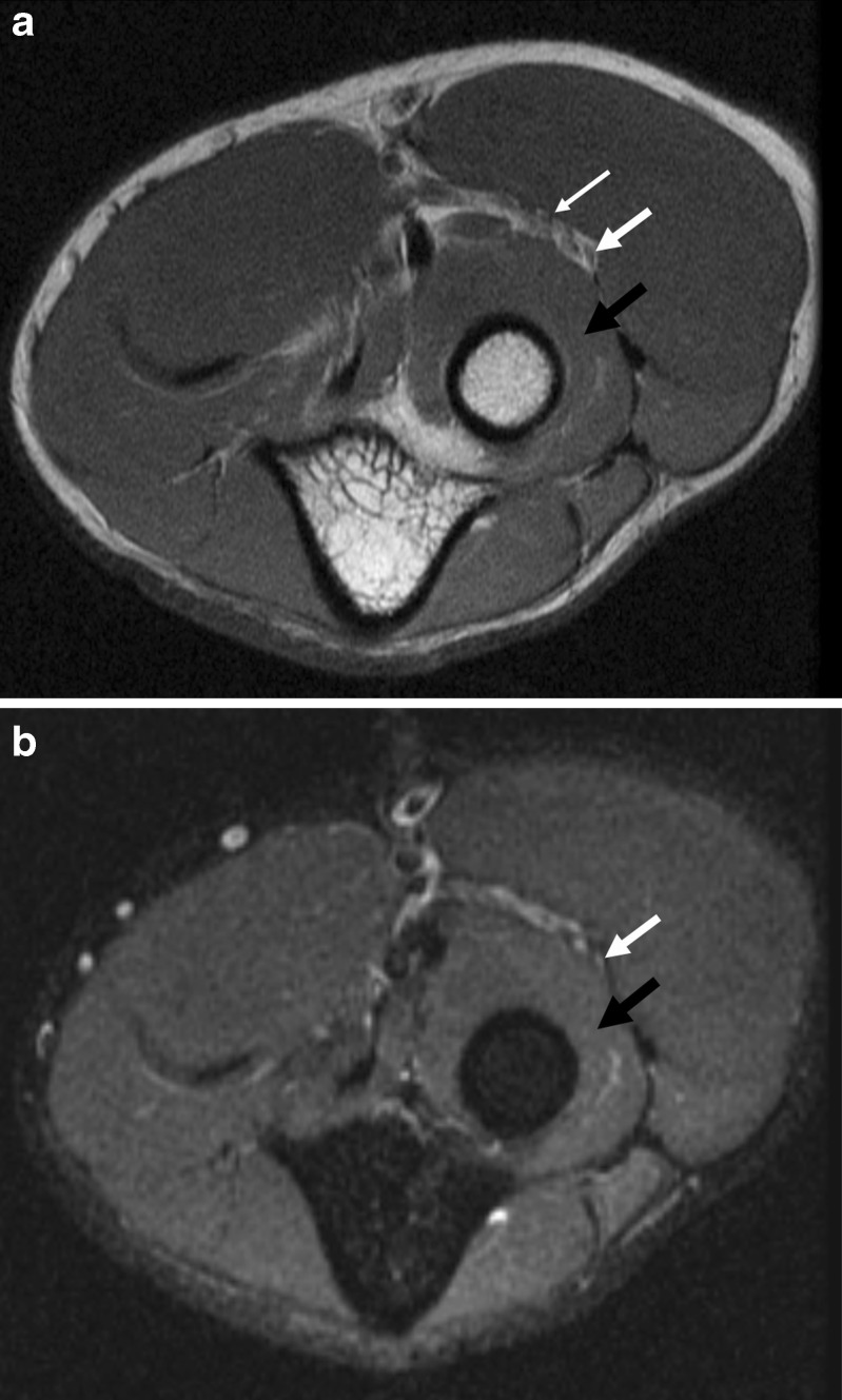
Fig. 1.
Axial fast spin echo (a) and inversion recovery (b) images of the left elbow demonstrate increased signal intensity in the posterior interosseous nerve (thick white arrow), without mass compression of the nerve. The superficial branch of the radial nerve (thin white arrow) has a normal signal and morphology. The supinator muscle has a normal signal, without evidence of a denervation effect (black arrow)

